A University of Iowa Technology Institute graduate research assistant continues to gain recognition as he works to make magnetic resonance imaging (MRI) processes quicker and more effective.

Aniket Pramanik, a PhD candidate in computer engineering and a research assistant in ITI’s Computational Biomedical Imaging Group (CBIG), recently won the Summa Cum Laude Merit Award for a research proposal he presented at the International Society for Magnetic Resonance in Medicine 2021 annual meeting.
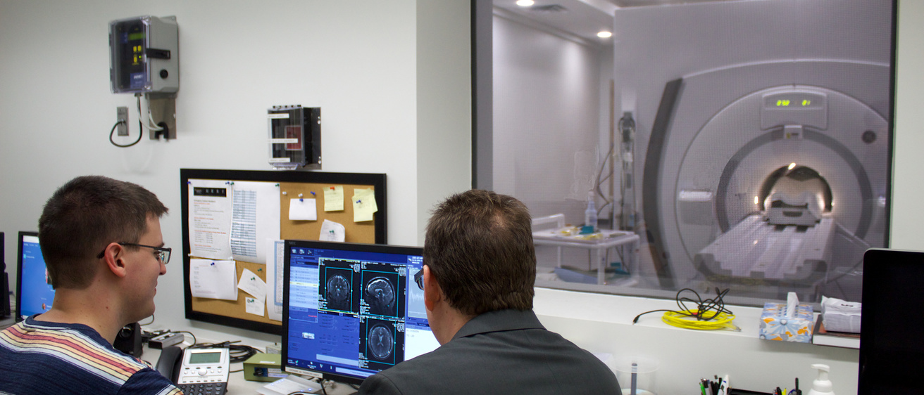
Pramanik proposed an algorithm using a new deep learning approach that would speed up MRI scan times while producing high-quality, deblurred images from under-sampled data that still allow segmentation of regions of the brain, an important task in medical image analysis.
“The reconstruction algorithm is effective in reducing MRI scan times, thus reducing discomfort for claustrophobic, obese, or older people and children,” Pramanik said.
The Summa Cum Laude Merit Award received approximately 5,000 abstracts vying for the distinction, and only the top 2% in terms of quality of research were chosen for the award. Granted only to first-author students, this award is highly competitive.
It is Pramanik’s latest award in a series of recognitions since beginning his thesis work three years ago. Attending his first conference in 2019, Pramanik presented a preliminary study at the International Symposium on Biomedical Imaging in Venice, Italy, and won the award for “Best Machine Learning Paper.”
Pramanik’s latest research proposal confronts pressing issues in MRI. Neurodegenerative diseases are characterized by the progressive degeneration of the structure and function of important brain regions. Volume estimates of certain brain regions serve as indicators for determining the severity of such diseases.
Magnetic resonance (MR) image segmentation of certain brain regions is vital for quantifying neurodegenerative disease progression. For example, the atrophy of hippocampal sub-regions and cortical thickness are established biomarkers for the progression of Alzheimer’s Disease. The volume estimates of these regions play important roles in the early diagnosis and prognosis of patients.
The issues arise in the amount of time it takes to achieve high-resolution images in normal scans, and in the lower quality of images that are produced by accelerating scans and collecting fewer data, a process known as undersampling. These imperfections restrict the accuracy of volume estimates of the brain’s subregions, leading to low sensitivity to and recognition of early disease-induced changes and long follow-up times to evaluate neurological disorder progression.
“The proposed research would lead to early diagnosis of neurodegenerative diseases in general, along with the estimation of their severity,” Pramanik said. “It would help doctors to plan an appropriate treatment for patients at an early stage that might help to cure the disease.”
The Computational Biomedical Imaging Group (CBIG) is among the more than 30 research labs and centers at the Iowa Technology Institute, a multidisciplinary research hub at the University of Iowa. It is grounded in engineering and a strong belief is science. CBIG is directed by Mathews Jacob, a professor of biomedical engineering.